On-line Developmental Atlas

The following developmental stages of the chick and pig embryos have been serially sectioned and labeled and unlabeled images of these sections have been compiled.
On-line Developmental Atlas |
|
|
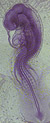
24-hour Chick Embryo |
| 33-hour Chick Embryo | |
| 48-hour Chick Embryo | |
| 72-hour Chick Embryo | |
|
|
| 10-mm Pig Embryo | |
The goal of the lab is to introduce you to a microscopic examination of the vertebrate embryo. Histological sections of the chick and the pig embryo are used to illustrate changes that occur as the embryo develops. Serial sections of these embryos have been scanned and can be visualized from any computer station on campus or via modem from a remote site 24 hours a day, seven days a week. This digital atlas is experimental.
You will be responsible for all labeled structures in the digital atlas.